|
Histology-World!
|
|
|
Histology Fact Sheet: Muscle |
||||||||
|
|
|
Muscle tissue is specialized for contraction. There are two chief categories of muscle: striated and non striated muscle (smooth muscle). Sometimes muscle is categorized as three types: skeletal, cardiac, and smooth. Striated MuscleStriated muscle can be sub-categorized into cardiac muscle and skeletal muscle. Both skeletal muscle and cardiac muscle have visible striations. Collectively, skeletal muscle and cardiac muscle are classified as "striated muscle". Skeletal Muscle
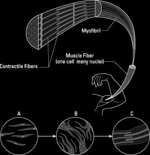
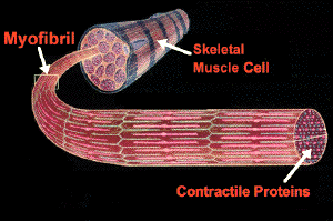
The basic unit of a muscle is the muscle fiber. Skeletal muscle fibers are multinucleated. On a histology slide, it can be seen that the nuclei are located on the periphery of the cell. These muscle fibers are striated. Cardiac Muscle
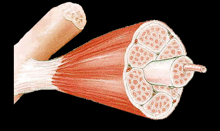
The heart is composed of cardiac muscle. Myocardium is the muscular layer of the heart. Thus, the myocardium is composed of cardiac muscle. Cardiac muscle is under control of the autonomic nervous system. The contraction of cardiac muscle is involuntary, strong, and rhythmical. The cardiac muscle cell is rectangular in shape. On a histology slide, it can be seen that the cardiac muscle cell has one central nucleus, like smooth muscle, but it also is striated, like skeletal muscle. In cardiac muscle, the nucleus is located centrally. Histology hint from Sarah Bellham: This is an important point, as both skeletal muscle and cardiac muscle are striated. The centrally placed nucleus seen in cardiac muscle is one of the things that can be used to distinguish between the two. On a histology slide, branching cells are seen in cardiac muscle. Histology hint from Sarah Bellham: this is an important point, as both skeletal muscle and cardiac muscle are striated. The branching seen with cardiac muscle is one of the things that can be used to distinguish between the two. When examining the histology it can be seen that occasionally, cardiac muscle is bi-nucleated. Intercalated discs are seen in cardiac muscle. Intercalated discs are specialized junctions between cardiac cells. Smooth MuscleNon striated muscle is also called smooth muscle. Smooth muscle is found in the walls of the hollow internal organs such as blood vessels, the gastrointestinal tract, bladder, and uterus. Smooth muscle is involuntary muscle. Smooth muscle is under control of the autonomic nervous system. Smooth muscle cannot be controlled consciously and thus acts involuntarily. Smooth muscle contracts slowly and rhythmically. Smooth muscle is composed of spindle shaped cells. In smooth muscle, there is also a centrally placed nucleus. Connective TissueThe outer connective tissue covering of a muscle is the epimysium. Within the muscle, there are subdivisions called fascicles. The perimysium surrounds these muscle fascicles. The endomysium is the covering around an individual muscle fiber. Histology hint from Sarah Bellham: The prefix "peri" means around, such as in the word "perimeter". The prefix "endo" means within or inner, such as in "endosteum","endocrine", "endoscope". Sarcolemma and SarcoplasmThe sarcolemma is the plasma membrane of a muscle cell. The sarcoplasm is the cytoplasm of a muscle cell. |
||
|
|
| Copyright (c) Histology-World and its licensors. All rights reserved. |
 Skeletal muscle is responsible for skeletal movement. The central nervous system (CNS) controls the skeletal muscles. Skeletal muscles are under conscious, or voluntary, control.
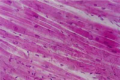
 Myofibril