|
Histology-World!
|
|
|
|
|
Key Histology FeaturesInstructions: Run your mouse over the histology slide want to view. Key histology features are described. If a link is present, click to view and listen to the histology audioslide.
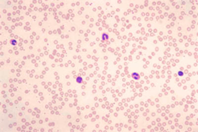
BloodThese are histology slides of a peripheral smear of blood. The majority of the cells are red blood cells. The red blood cells have a central area that is paler. This is called the "central pallor". This is seen because the red blood cells are biconcave discs. The diameter of a red blood cell is 7-8 microns. You can use a red blood cell as a ruler, to estimate the diameter of the other cells, provided the RBC are all uniform in size, as they are here. Five white blood cells can be seen histology slides (1) and (4). The white blood cells are the larger cells with dark nucleus. The platelets look like cellular debris in this histology slide (1).
|
| Copyright (c) Histology-World and its licensors. All rights reserved. |