|
|
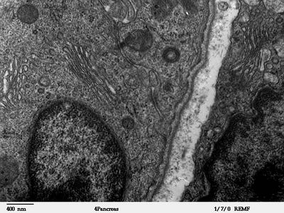
Desmosomes and Golgi - TEM
|
Transmission electron microscope image of a thin section cut through an area of mammalian pancreatic tissue. Image shows pancreatic cells, with nice examples of golgi and desmosomes (junctions). JEOL 100CX TEM
Image courtesy of Louisa Howard.
|
|