|
Histology-World!
|
|
|
|
|
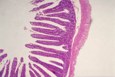
Key Histology FeaturesInstructions: Run your mouse over the histology slide want to view. Key histology features are described. If a link is present, click to view and listen to the histology audioslide. Small IntestineThe small intestine has a mucosa, submucosa, muscularis externa, and serosa. On these histology slides (1), (2), (3), and (4), note the prominent villi. The apical surface has finger like projections (villi). This is in contrast to the large intestine. There are goblet cells on the villi. These are the clear looking hollowed out spaces. On histology slides (4) and (5), the brush border is visible. Histology hint from Sarah Bellham: Compare both the apical surface and the number of goblet cells in the small intestine to the apical surface and number of goblet cells in the large intestine.
|
| Copyright (c) Histology-World and its licensors. All rights reserved. |